Ophthalmology
Capital Clinic Riga Ophthalmologists provides consultations, examinations, diagnostics, conservative treatment and refraction correction. They perform an in-depth investigation and conservative treatment of glaucoma patients, diagnostics of eye complications related to diabetes as well as diagnostics and treatment of retinal diseases.
The first visit to the Ophthalmologist
49 EUR
Everybody, starting from infancy, should get regular eye examinations. Preventive eye examinations for children are recommended at the age between 13 – 24 months, then at the age between 3 – 4 years and before attending the school – at the age between 6 – 7 years or any other time, if recommended by the eye doctor. Adults should get their eyes examined once a year after the age of 40 or any other time, if recommended by the eye doctor. Ophthalmologist should be visited whenever having some complaints about the vision or eye health – if the vision has worsened or blurred, if distortion of lines or image is observed, eyes are inflamed, foreign bogy gets in the eye and other similar cases.
The ophthalmology office in Capital Clinic Riga is equipped with full range state of the art up to date technologies for accurate evaluation of eye health and vision diagnostics and, therefore, providing the most effective treatment possible:
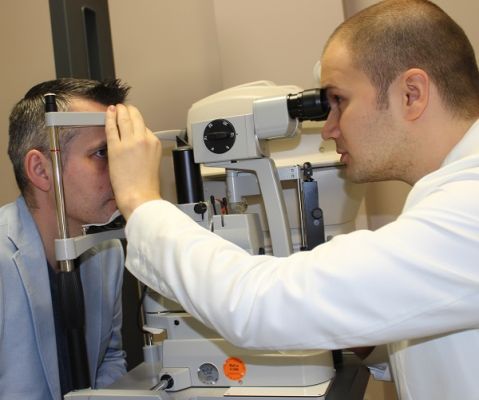
Autorefractor Keratometer

Autorefractor keratometer is the first device used by the eye doctor to examine your eyes when visiting a modern eye doctor’s office. It automatically detects the optical power of the eye, corneal curvature and its regularity. It helps to determine the strength of the lens needed to correct the optical aberrations or the optical errors of the eye. It also helps to assess the general condition of the outer parts of the eye. If necessary it also provides the necessary information for prescription of the lenses.
Subjective Refraction Determination

During this examination the eye doctor determines your visual acuity and the best corrected visual acuity for each eye separately. This is done my reading the table of letters, numbers or symbols displayed on a screen. If the visual acuity does not meet the standards, visual acuity correction is made using corrective lenses placed in a special frame.
Contactless Tonometer with Pachymeter

This is a contactless device for measuring eye pressure. It is combined with a built-in pachymeter which automatically measures corneal thickness.
The eye pressure reading obtained using tonometry method by its own is approximate, because patients with thicker cornea will have lower pressure reading and vice versa – patients with thin cornea will have higher pressure reading. In Capital Clinic Riga eye cabinet it is possible to make an accurate eye pressure measurement in correlation with pachymetry (corneal thickness) data.
Dioptometer

Dioptometer carries out a complete analysis of eyeglass lenses. The patient's current glasses are placed in the device and it determines their optical strength. It is possible to analyze monofocal (spherical and cylindrical), bifocal and progressive eyeglass lenses. Using the data obtained from this examination, the eye doctor assesses if the glasses are suitable for the patient and, if necessary, prescribes new ones.
Optical Coherence Tomography (OCT)

OCT is a modern medical imaging method that eye doctor uses to make high resolution eye tissue analysis, instantly receiving cross-sectional analysis of the retina, optic nerve, cornea and anterior chamber angle. It is also possible to make photo documentation of the retina. This device also helps to analyze the characteristics of retinal surface, structure of the retina, retinal nerve fiber layer thickness and carries out a structural analysis of the optic nerve. The examination is painless, with no contact with the surface of the eye and safe for patients’ health.
This examination is invaluable when diagnosing glaucoma patients. It is essential to measure retinal nerve fiber layer thickness around the optic nerve using OCT. This is very important because reduced RNFL (retinal nerve fiber layer) is considered an early sign of glaucoma. With OCT it is possible to diagnose glaucoma earlier that with the gold standard method in glaucoma diagnostics – automated visual field test (perimerty). Automated visual filed test shows pathological changes only when the patient has lost a large amount of optic nerve fibers.
OCT is used for diagnostics of eye diseases, for following the dynamics of eye diseases when they are progressing, as well as for evaluating the effectiveness of a certain treatment.
Automated Visual Field Test (Perimetry)

With OCT it is possible to diagnose structural optic nerve damage, but with the help of automated visual field test functional defects can be found. Automated visual field test is essential for glaucoma diagnostics, evaluation of disease progression and treatment effectiveness assessment.
Normal visual field covers 90 degrees temporally (to the side), 60 degrees nasally (to the side of the nose) and upwards and 70 degrees downwards, when fixing the view in one point. With this examination method the eye doctor assesses whether patients’ visual field parameters matches the ones that are relevant for the patients’ age group.
The examination involves the view fixation in one point. Light impulses are projected in patient’s visual field. When patient sees a light impulse he should press a button that is connected with computer system. Light impulses are projected in different areas of the visual field, but the patient does not know the next test point location. Also the intensity of light stimulus is variable. This way absolute or relative visual field defects can be diagnosed.
Automated visual field analyzer statistically processed data, in addition evaluating the reliability of the test criteria. Visual field defects are significant in assessing glaucoma progression and often help in diagnosing neurological abnormalities. Visual field test is also carried out for people who want to receive their driver’s license.
Eye Ultrasound

It is an important diagnostic device in ophthalmology. Eye ultrasound device helps to get instant two-dimensional images of eye tissue and information about the state of the intra-orbital tissue. This examination is painless and safe for patient’s health.
Ultrasound can be used very widely, especially in cases where the optical media of the eye is not clear. This can include clouding of the lens, corneal pathology and vitreous haemorrhage. In these situations the deeper structures of the eye cannot be visualized by other diagnostic methods. The most common indications for ultrasound usage include retinal detachment, intra-ocular formation localization, extra-ocular muscle thickness measurement, and vitreous opacities diagnostics.
To apply for a consultation and learn more, please, call:
66 333333, 29334224, e-mail us at: info@capitalclinicriga.lv
or use e-appointment >>